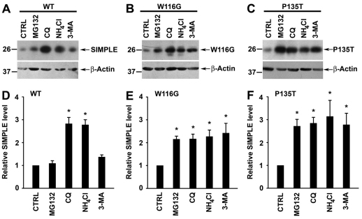
Fig. 8.
Clearance of CMT1C-associated mutant SIMPLE proteins by both the proteasome and autophagy pathways. (A–C) Steady-state protein levels of WT (A), W116G (B), or P135T (C) SIMPLE in HEK293 cells treated with 20 μM MG132, 100 μM chloroquine (CQ), 50 μM NH4Cl, 10 mM 3-methyladenine (3-MA) or vehicle (CTRL) were analyzed by immunoblotting with antibodies against the Myc tag and β-actin. (D–F) The relative level of WT (D), W116G (E), or P135T (F) SIMPLE protein in the proteolysis inhibitor-treated cell lysates was normalized to the β-actin level and expressed relative to the normalized SIMPLE protein level in the corresponding vehicle-treated control lysate. Data represent mean ± s.e.m. from at least three independent experiments. *P<0.05 compared with vehicle-treated control.

