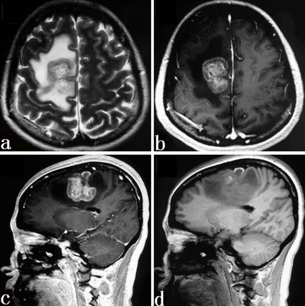
Figure 1.

Axial T2-weighted MR image (a) showing a mass with mixed signal intensity and a surrounding edema area. On the T1-weighted image after the administration of contrast material (b and c), the mass has an inhomogeneous enhancement. Sagittal T1-weighted MR image without contrast (d) depicting a striped hemorrhage.
