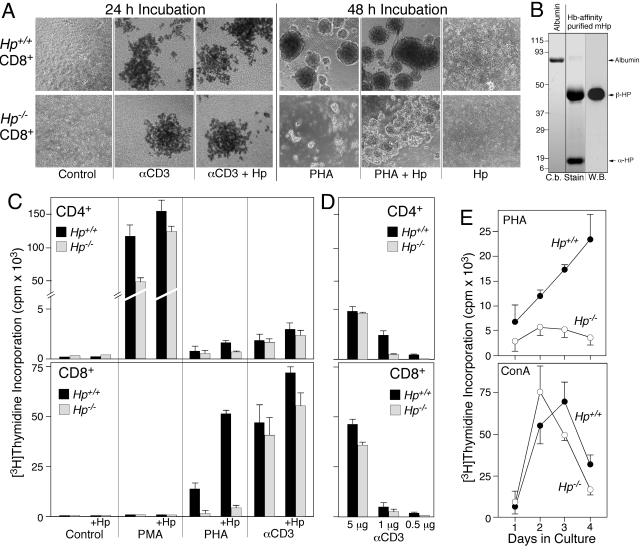
Figure 7.
T cells from Hp-deficient mice show an altered response to mitogens. T cells from Hp-deficient mice show an altered response to mitogens (A), microphotographs (4×) of purified CD8 cells isolated from Hp+/+ and Hp−/− C57BL/6J mice treated for 24 h with anti-αCD3 (αCD3), or for 48 h with PHA. Selected cultures also included 1 mg/ml mouse Hp. (B) Coomassie blue-stained SDS-PAGE gel (C.b. Stain) demonstrates purity of Hp (8 μg) isolated from acute phase serum of Hp+/+ C57BL/6J. A duplicate lane was used for Western blotting (W.B.) reacted with rabbit anti-Hp (recognizes only epitopes in the β-subunit). (C and D) [3H]Thymidine incorporation was determined in cultures of purified CD4 and CD8 cells that had been maintained in the presence of the indicated mitogen for 72 h. (E) DNA synthesis was determined in nonfractionated splenocytes from Hp+/+ and Hp−/− mice as a function of duration in culture in the presence of PHA or Con A.

