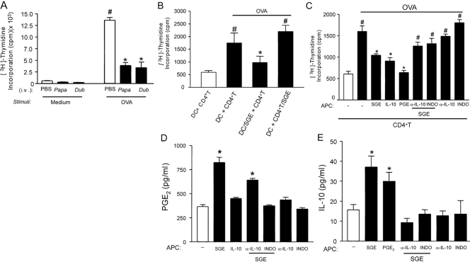
Figure 5.
The suppressive effect of SGE on OVA-induced lymphoproliferation depends on PGE2 and IL-10. OVA-immunized and sham-immunized mice (Control) were i.v.-treated with PBS or SGE from P. papatasi or P. duboscqi vector (one salivary gland/i.v./animal) 48 h before splenocyte harvesting. (A) Splenocytes were cultured with or without OVA (10 μg/mL) during 72 h, and the proliferation assay was performed by [3H]thymidine incorporation. (B) CD4+T or DC (1×10⁶ cells/ml) from OVA-immunized and naïve mice, respectively, were incubated overnight with or without P. papatasii SGE (0.5 gland), and coculture of CD4+T and DC (10:1) was performed for 72 h in the presence of OVA (same dose). In another set of experiments, DC (1×10⁶ cells) from naïve mice were incubated overnight with SGE (0.5 gland), IL-10 (100 μg/ml), PGE2 (1 μM), antibody against IL-10 (α-IL-10; 10 μg/ml), or indomethacin (10 μg/ml). α-IL-10 or indomethacin was also added in the presence or absence of SGE. Afterwards, CD4+T cells of OVA-immunized mice were added, followed by stimulation with or without OVA. The lymphoproliferation was assessed by [3H]thymidine incorporation (C), PGE2 by RIA (D), and IL-10 by ELISA (E). The results are expressed as the mean ± sem of at least two separate experiments made in triplicate (n=3 per group); #, P < 0.05, compared with the medium stimulus; *, P < 0.05, compared with the saliva control (PBS) and DC + CD4+T groups.

