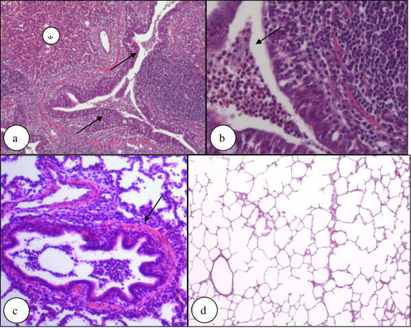
Figure 4.
Histological evaluation of lung samples. Figure 4a-b show lung tissue from a pig treated with NCT showing atelectases (*) and infiltration of the bronchial and alveolar system by neutrophilic granulocytes that are present in the lumen of a bronchial airway (arrows) (hematoxylin-eosin staining; original magnification 100× (a) and 400 × (b); p > 0.1 for frequency of occurrence between test and control group). Figure 4c shows a mild increase of thickness of the bronchial wall on serial sections, although this finding was patchy in the same lung and not uniform in the same group (arrow) (hematoxylin-eosin staining; original magnification 200×). Figure 4d shows lung tissue from a pig treated with NaCl showing neither atelectasis nor neutrophilic granulocytic infiltration (hematoxylin-eosin staining, original magnification 50x; p > 0.1 for frequency of occurrence between test and control group).

