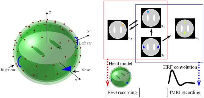
Figure 3. Head model and the construction of synthetic data.
(a) Head model: 2452 voxels within a concentric three-sphere head model with 64 electrodes on the upper surface. The two holes in source slice are ‘white matter.’ (b) A schematic illustration of the procedure to generate simulated time series. The spatial profile of each source is drawn with different color and the background is shown in gray and white. EEG signals are recorded from S1, S2 and S3 (red-bordered areas), which generate scalp potentials through the head model. S1, S3 and S4 (blue-bordered areas) are filtered by convolution with a gamma function to yield fMRI recording. S2 and S4 are assumed to be fMRI and EEG blind respectively.

