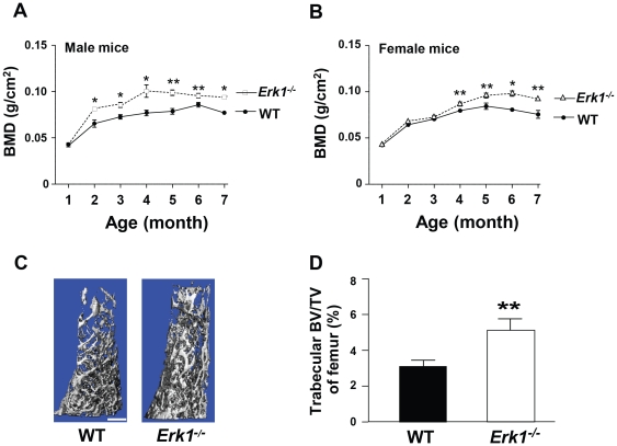
Figure 5. Erk1−/− mice have increased BMD and BV/TV.
BMD of age and sex matched Erk1−/− and WT mice was measured from birth to 7 months of age. The BMD of male (A) and female (B) WT and Erk1−/− mice is shown (N = 5 mice in each group). (C) Representative μCT reconstructions of WT and Erk1−/− femurs are shown. Scale bar = 1 mm. (D) Quantitative data comparing the left femur BV/TV between WT and Erk1−/− mice.

