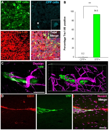
Figure 3. Histological evaluation reveals tumors contained cancer stem cells and their descendants that had association with blood vessels.
Tumors from the cell mixing experiments (n = 3) were evaluated to determine their composition. Subsequent evaluation of resulting tumors demonstrates that the majority of the cells within the tumor mass was of human origin and derived from CSC as confirmed by Tra-1-85 staining and YFP expression, shown in representative micrographs (A) and bar graph (B). Peripheral transplanted tumor cells (YFP positive CSCs and their descendants) were observed to have an association with blood vessels. Micrograph from multiphoton imaging and three-dimensional reconstruction (C) depict close association of tumor cells (green) with adjacent blood vessel (purple, illuminated by fluorescent dextran injection into the circulation prior to imaging). Histological examination of resulting tumors confirms close association of peripheral tumor cells to the vasculature using CD31 immunostaining (D; CD31 in red, tumor cells in green, nuclei in purple). Scale bar represents 50 µm. Data displayed as mean values +/- S.E.M. ***, p<0.001 as assessed by one-way analysis of variance (ANOVA).

