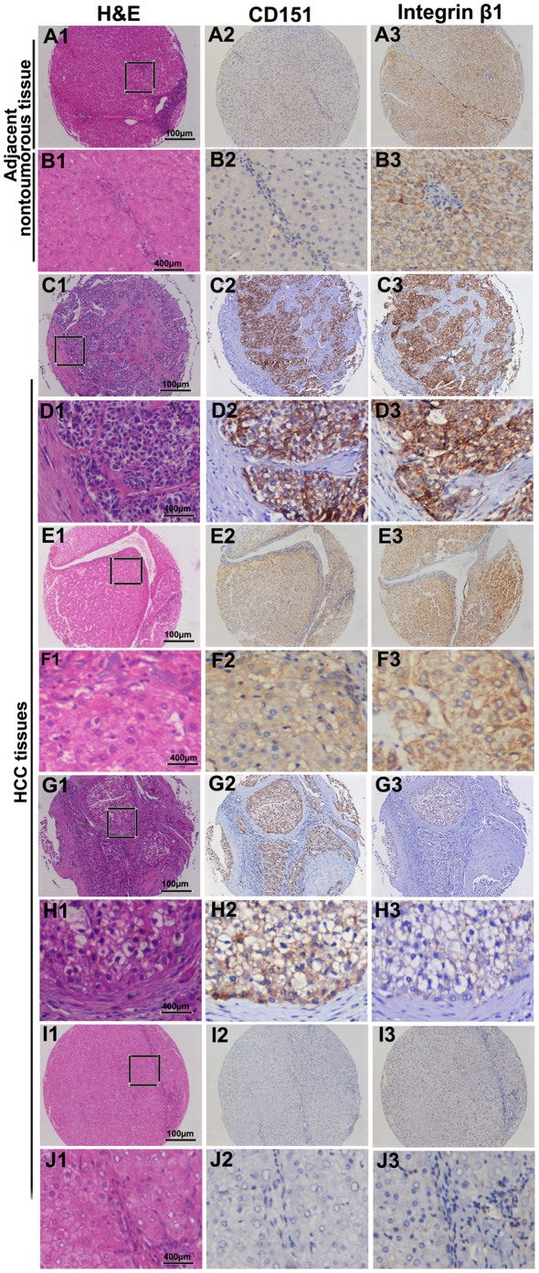
Figure 4. Expression of CD151/integrin β1 complex in HCC patients by immunohistochemistry.
After identification by H&E staining (A1, B1, C1, D1, E1, F1, G1, H1, I1, and J1), CD151 was mostly located on the membrane of tumor cells, with comparable variation (A2, B2, C2, D2, E2, F2, G2, H2, I2, and J2) and strong integrin β1 expression in most HCC cells, stroma fibroblasts, and epithelial cells of the bile duct (A3, B3, C3, D3, E3, F3, G3, H3, I3, and J3). A and B refer to adjacent nontumoral tissue. Representative tumor tissues are shown :C and D high expression of CD151 and integrin β1, E and F high expression of CD151 and low expression of integrin β1, G and H high expression of integrin β1 and low expression of CD151, I and J low expression of CD151 and integrin β1.

