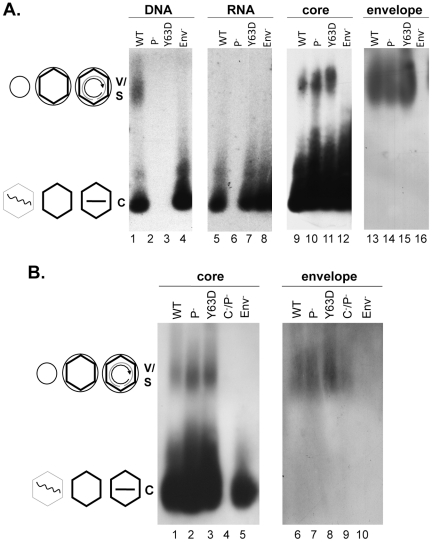
Figure 2. Analyses of HBV virion secretion by native agarose gel electrophoresis.
WT or mutant HBV genomes (Pol-, defective in pgRNA packaging; Y63D, competent in pgRNA packaging but defective in DNA synthesis; Env-, envelope deletion; C-P-, deficient for both core and polymerase expression) were transfected into HepG-2 cells. Viral particles concentrated from the culture medium were resolved by agarose gel electrophoresis and transferred to nitrocellulose membrane. Viral DNA (A, lanes 1–4) or core (A, lanes 9–12; B, lanes 1–5) and envelope proteins (A, lanes 13–16; B, lanes 6–10) were detected as in Figure 1 . Viral RNA (A, lanes 5–8) was detected as for viral DNA, except that the samples were resolved on a separate gel that was not treated with NaOH prior to transfer and the membrane probed with a plus-strand specific riboprobe. V, virions, containing either RC DNA or empty; C, naked capsids, containing DNA, RNA, or empty. The diagrams on the side depict the structures of the virions and capsids as described in Figure 1 .

