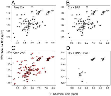
Figure 3. -1H-15N HSQC spectra of Crx homeodomain.
(A) 30 µM free 15N-labeled Crx. (B) 30 µM free 15N-labeled Crx plus 200 µM BAF2. (C) 30 µM free 15N-labeled Crx plus 30 µM 16 mer DNA (black). The spectrum in the absence of DNA is superimposed in red. (D) 30 µM free 15N-labeled Crx plus 200 µM BAF2 and 30 mM 16 mer DNA.

