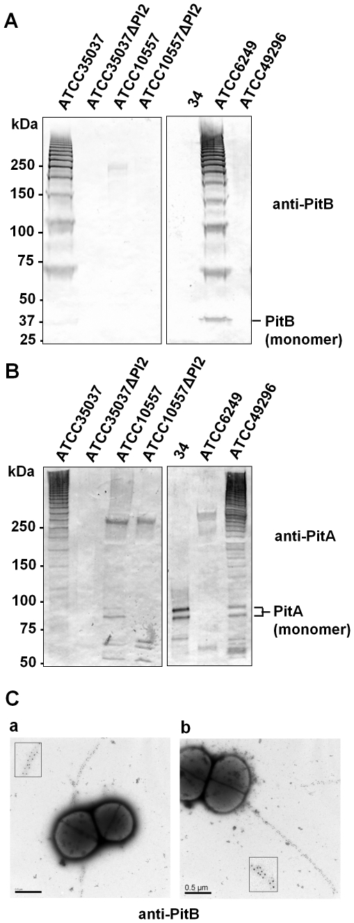
Figure 3. Detection of PI-2 pili on the surface of Mitis group streptococci.
Western blots with cell wall extracts of the indicated strains detected with anti-PitB antiserum (A) or anti-PitA antiserum (B). Both antisera were developed against the respective S. oralis ATCC35037 proteins. The positions of the PitB and PitA monomers are indicated. (C) Electron micrographs of immmunogold-labeled S. oralis ATCC35037 (a) and S. mitis ATCC6249 (b) using anti-PitB antiserum. Scale bars 0.5 µm.

