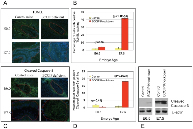
Figure 4. Apoptosis in BCCIP deficient embryos.
At days E6.5 and E7.5, embryo tissue sections were prepared to stain for apoptosis markers. TUNEL (panel A) and anti-cleaved caspase-3 (panel C) staining were performed on embryonic tissue sections at days E6.5 and E7.5 (indicated on the left of the panels). At this stage, the surrounding the embryos are the decidual tissues that normally undergo apoptosis, and the embryonic tissues are marked with red lines. The percentage of apoptotic cells are shown in panel B and D. Panel E is a Western blot showing the increase of cleaved caspase-3 in the BCCIP deficient embryos.

