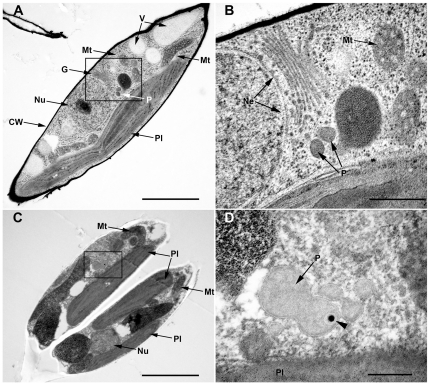
Figure 2. Immunolocalization studies of a peroxisomal GFP-fusion protein in P. tricornutum.
(A) Ultrathin section of P. tricornutum expressing Pex10-GFP in Epon without antibody labeling. The boxed area is shown in (B) at higher magnification and illustrates two peroxisomes in proximity to the nucleus, the golgi and the plastid. (C) Immuno labeling of Pex10-GFP in a dividing P. tricornutum cell. (D) higher magnification. The 20 nm gold particle, coupled to the secondary antibody is visible within the peroxisome (arrow head), which is surrounded by a lipid bilayer. CW, cell wall; G, golgi apparatus; Mt, mitochondrium; Ne, nuclear envelope; Nu, nucleus; P, peroxisome; Pl, plastid; V, vacuole; arrow head, 20 nm gold. Scalebars represent 2 µm (A, C), 500 nm (B) and 200 nm (D).

