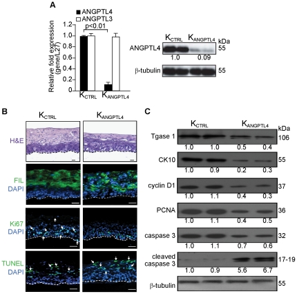
Figure 2. Organotypic skin coculture (OTC) of ANGPTL4-deficient human primary keratinocytes displayed impaired epidermal differentiation.
(A) Relative mRNA and/or protein levels of ANGPTL4 and ANGPTL3 in human primary keratinocytes transduced with either scrambled control (KCTRL) or ANGPTL4 siRNA (KANGPTL4). Values below immunoblot bands represent the mean fold differences in protein expression level when compared with KCTRL from 3 independent experiments. (B) Haematoxylin and eosin (H&E) and immunofluorescence staining of OTC sections constructed with either control (KCTRL) or ANGPTL4-knockdown (KANGPTL4) human keratinocytes. Late (fillaggrin, FIL) differentiation markers, proliferating (Ki67) and apoptotic (TUNEL) cells were identified using indicated antibodies or assay. White dotted lines indicated epidermis-dermis junctions. Sections were counterstained with DAPI (blue). Scale bars represent 40 µm. (C) Representative immunoblot of early epidermal differentiation (cytokeratin 10, CK10), terminal differentiation (transglutaminase type I, Tgase 1), proliferation (PCNA and cyclin D1), and apoptosis (cleaved caspase 3) markers in isolated epidermis of indicated OTCs. All immunoblot data are from three independent experiments performed in duplicate. β-tubulin serves as a loading and transfer control.

