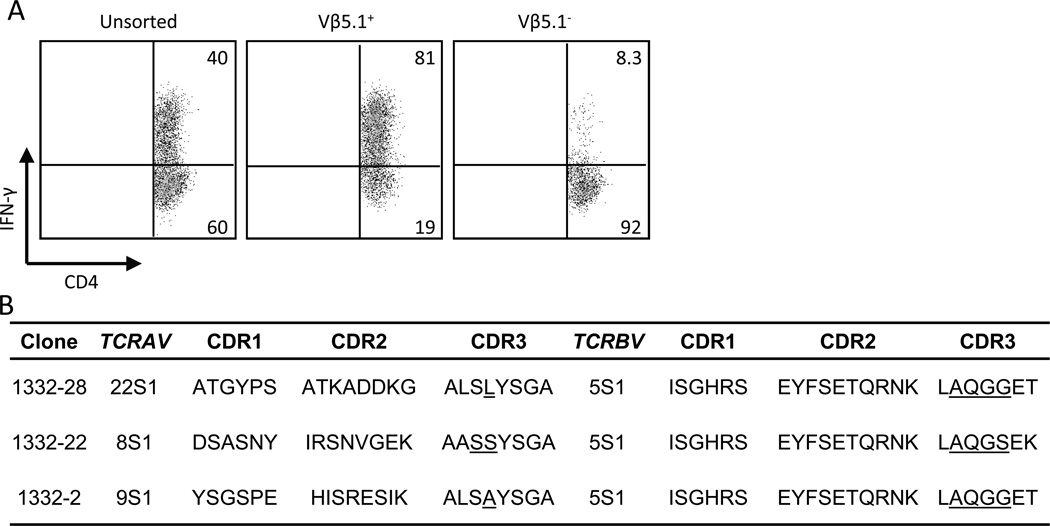
FIGURE 1.
Deduced amino acid sequences of a set of Be-responsive, Vβ5.1+CD4+ T cells. A, Intracellular IFN-γ staining of a Be-responsive T cell line derived from the BAL of a CBD patient is shown. The cells were stimulated with either medium alone or BeSO4 and subsequently stained for CD4, Vβ5.1 and intracellular IFN-γ expression. The number in the upper right quadrant of each density plot indicates the percentage of CD4+ T cells expressing IFN-γ. B, Analysis of deduced TCRA and TCRB CDR1, CDR2 and CDR3 amino acid sequences expressed by the Be-responsive T cell clones derived from the T cell line in A. Underlined amino acids indicate those encoded by non-germline nucleotides. These sequence data are available from GenBank under accession numbers JF834314 - JF834319 http://www.ncbi.nlm.nih.gov/genbank/.

