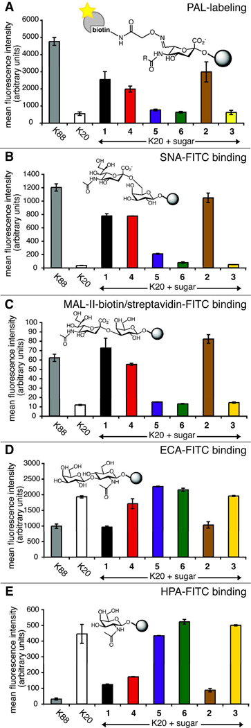
Figure 4. Cell surface sialic acid measured by flow cytometry.
BJA-B K20 cells were grown in serum-free media supplemented with 100 µM 1–6. (A) Total sialic acid detected by PAL; (B) Siaα2–6 galactose (Siaα2–6Gal) epitope detected by SNA lectin; (C) Siaα2–3Gal epitope detected by MAL-II lectin; (D) unsialylated terminal N-acetyllactosamine (LacNAc) detected by ECA lectin; (E) unsialylated terminal N-acetylgalactosamine (GalNAc) detected by HPA lectin. Error bars represent the standard deviation for three technical replicates.

