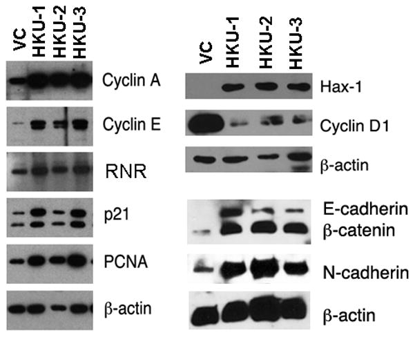
Figure 4. Analysis of proteins associated with cell cycle progression.

Cell extracts of stable clones (20 μg of total protein for cyclin A, cyclin E, RNR and PCNA; 5 μg for Hax-1, cyclin D1, E-cadherin, N-cadherin and β-catenin) were analyzed by Western blot using respective antibodies. β-actin was used as loading control. Fig. 1B Sam68 blot was reused for Hax-1 and cyclin D1 expressions.
