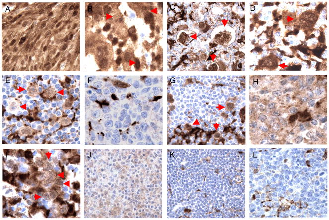
Figure 2.
Cases of follicular dendritic cell sarcoma (A), histiocytic sarcoma (B), classical Hodgkin lymphoma (C; D), nodular lymphocyte predominant Hodgkin lymphoma (E), diffuse large B cell lymphoma, not otherwise specified (F), T cell/histiocyte rich large B cell lymphoma (G), primary mediastinal (thymic) large B cell lymphoma (H, I), extranodal marginal zone lymphoma (J), small lymphocytic lymphoma (K), and Burkitt lymphoma (L) showing positive (A–E; G–J) and negative (F, K; L) staining for TNFAIP2 in tumor cells (red arrows in B–E, G; I). All images were photographed at 1000×.

