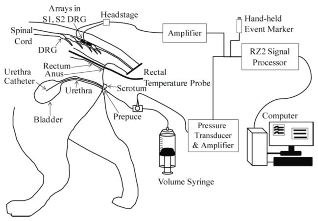
Figure 1.
Experimental setup. A urethral catheter was inserted for access to the bladder for fluid infusion and pressure monitoring. A 90-channel microelectrode array (Blackrock ICS-96 MultiPort split array: 50 and 40 electrodes) was inserted in the S1 and S2 DRG on one side. Sensory stimuli were applied by changing the bladder volume, moving the urethra catheter, pinching the prepuce, brushing the scrotum, moving the rectal temperature probe and brushing around the anus. Neural signals from the microelectrodes and the bladder pressure were viewed and stored on a PC after processing with a TDT RZ2 system.

