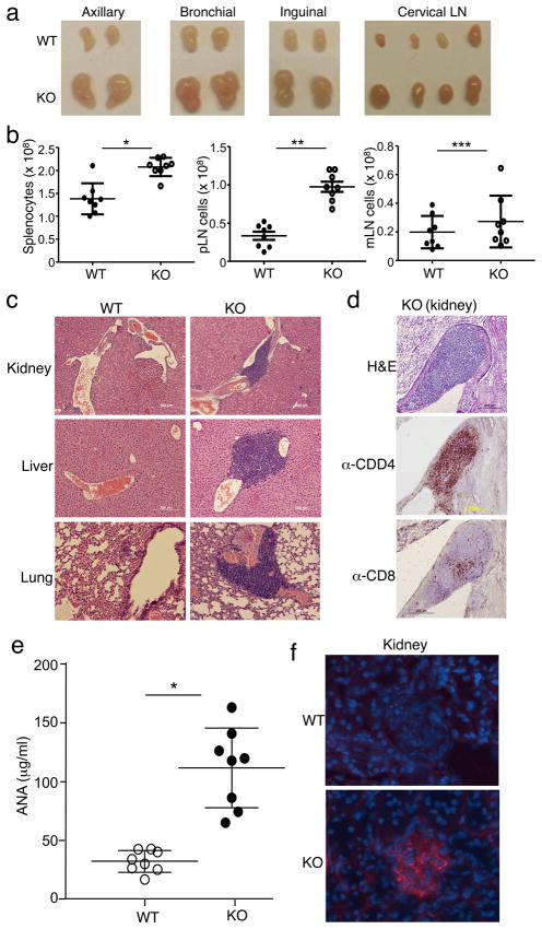
Figure 4. Peli1−/− mice spontaneously develop autoimmune symptoms.
(a) Lymph nodes isolated from age- and sex-matched Wild-type (WT) and Peli1−/− (KO) mice (10 week old). (b) Total cell numbers were counted in the spleen, peripheral lymph nodes (pLN), and mesentery lymph nodes (mLN) of 6 month old mice. Data are presented as mean value of multiple mice (each circle represents one mouse). *P<0.0002, **P<0.0001, and ***p=0.3457 (two-tailed unpaired t test). (c) Hematoxylin-eosin staining of the indicated tissue sections from 6 month old WILD-TYPE and KO mice, showing immune cell infiltrations. Original magnification, 100x (kidney) and 200x (liver and lung). (d) Kidney tissue sections were subjected to either hematoxylin-eosin staining or immunohistochemistry using anti-CD4 or anti-CD8 antibodies. Original magnification, 20x. Data in c and d are representative of multiple mice. (e) ANA ELISA of sera collected from age-matched wild-type and Peli1−/− mice (8 each; 10 to 24 month old). *P<0.001 (two-tailed unpaired t test). (f) Immunofluorescence staining of kidney tissue sections of wild-type and Peli1−/− mice to detect immune-complex deposits (red). Glomeruli were visualized by DAPI counterstaining (blue). Original magnification, 40x. Data are representative of 4 wild-type and 4 Peli1−/− mice.

