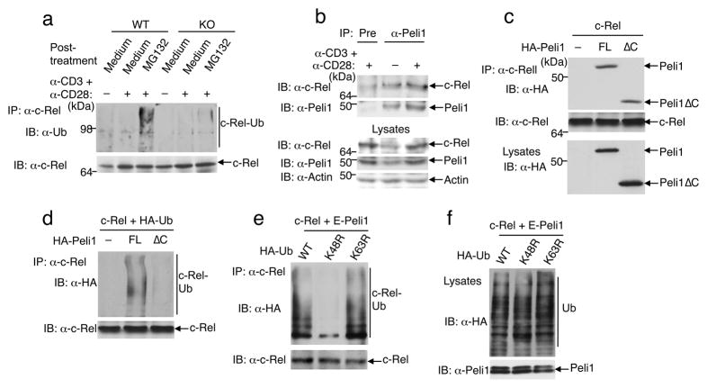
Figure 7. Peli1 induces c-Rel ubiquitination.
(a) Wild-type (WT) and Peli1−/− (KO) T cells were incubated for 16 h in the absence (−) or presence (+) of anti-CD3 plus anti-CD28 and then further incubated for 6 h with medium or MG132. Cell lysates were subjected to c-Rel IP, followed by IB. (b) Wildtype T cells were not treated (−) or stimulated (+) with anti-CD3 plus anti-CD28 for 16 h. Cell lysates were subjected to c-Rel-Peli1ΔcoIP (top two panels) or direct IB (bottom three panels). Data in a and b are representative of 3 independent experiments performed using multiple mice. (c) HEK293 cells were transfected with c-Rel along with full-length (FL) Peli1 or Peli1ΔC. Total cell lysates were subjected to c-Rel-Peli1ΔcoIP (top two panels) or Peli1 IB (bottom panel). (d) 293 cells were transfected as indicated. Immunoprecipitated c-Rel was subjected to IB to detect its conjugation with HA-ubiquitin and its expression level. (e, f) 293 cells were transfected with c-Rel and Etag-Peli1 along with HA-tagged wild-type (WT) or mutant forms of ubiquitin. Cell lysates were subjected to c-Rel ubiquitination (e) or direct IB (f) assays. Data in c-f are representative of 3 independent experiments.

