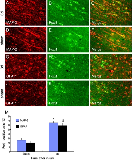
Fig. 3.
Double immunofluorescence staining for Foxj1 and different phenotype-specific markers in adult rat brain 3 days after TBI. The sections from sham and injured brains 3 days after TBI were immunostained with Foxj1 (green, b, e, h, k) and different cell makers, such as MAP-2 (a marker of neurons, red, a, d) and GFAP (a marker of astrocytes, red, g, j), and the co-localization of Foxj1 with different phenotype-specific markers were visualized in the merged images (c, f, i, l). a–c, g–i Immunostaining for Foxj1 with MAP-2 and GFAP at 3 days after TBI; d–f, j–l Immunostaining for Foxj1 with MAP-2 and GFAP in sham brain. m Quantitative analysis of different phenotype-specific markers positive cells expressing Foxj1 (%) in sham group and 3 days after injury. The change of Foxj1 was striking in neurons and astrocytes; *, # indicate significant difference at P < 0.05 compared with the sham group. Error bars SEM. Scale bars 20 μm (a–l)

