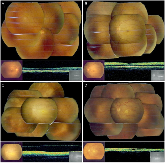
Fig. 2.
(A) The fundus photo of the right eye shows no abnormal findings. (B) At 3 hours after the autologous fat injection, a photo of the fundus of the left eye shows a cherry red spot with visible emboli in several retinal arteries. (C) At 24 hours after the injection, a photo of the fundus of the left eye shows marked retinal edema, disc swelling and multiple fat emboli. (D) At 2 months after the injection, a photo of the fundus of the left eye shows optic disc atrophy, multiple retinal hemorrhages and a fibrous change on its posterior pole.

