
Fig. 6.
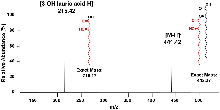
MS3 spectrum of lipid A isolated from E. coli MLK1067 expressing Vc0212. The doubly deprotonated lipid A ion (m/z 891.5) from MLK1067 expressing Vc0212 was subjected to 193 nm UVPD (see Fig. S4) followed by CID of the fragment ion of m/z 441.4. The ion of m/z 441.4 is interpreted as the deprotonated intact 3′-acyloxyacyl moiety of lipid A (exact mass calculated to be 442.37 u, proposed structure shown in the spectrum). The MS3 fragment ion of m/z 215.42 is interpreted as deprotonated 3-hydroxylauric acid (exact mass 216.17 u, proposed structure shown in the spectrum).