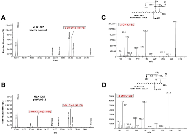
Fig. 7.
Gas chromatography/mass spectrometry (GC/MS) analysis of hydroxylated fatty acids from LPS isolated from E. coli MLK1067 expressing Vc0212. LPS was purified from strain MLK1067, hydrolysed, and converted to TMS derivatives to generate fatty acid methyl esters. (A) shows the total ion chromatogram (TIC) of the GC/MS analysis of hydroxy fatty acids from MLK1067 containing the vector control showing the presence of TMS-3-hydroxymyristoylmethylester (retention time: 30.172). (B) shows the TIC of the GC/MS analysis for MLK1067 containing pWVc0212 showing the presence of both TMS-3-hydroxymyristoylmethylester (retention time: 30.171) and TMS-3-hydroxylauroylmethylester (retention time: 21.564). (C) and (D) show the EI mass spectra of peaks at 30.171 and 21.564 found in the TIC of MLK1067/pWVc0212. Based upon fragmentation patterns of TMS derivatives of bacterial acid methylester standards (Fig. S5), electron ionization/mass spectrometry analysis of the 30.171 peak was indicative of a TMS-3-hydroxymyristoylmethylester (C) and analysis of the 21.564 peak indicative of TMS-3-lauroylmethylester (D). The EI mass spectra of the peak at 30.172 found in the TIC of MLK1067 vector control (A) was identical to that shown in (C). The key cleavages, indicated by dashed lines, are shown for each inserted structure. Sugars present in the sample are also indicated.

