
Figure 2.
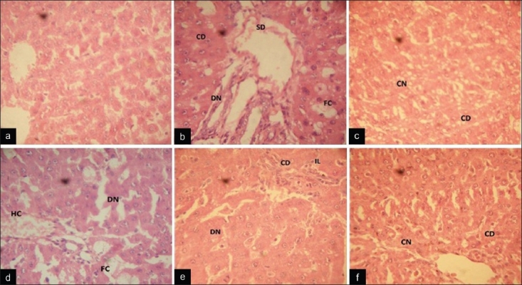
Photomicrograph of paraffin-embedded rat liver sections stained with hematoxylin and eosin. (a) Liver section of a normal control rat liver showing normal architecture with no cellular degeneration and necrosis. (b) Liver sections of CCl4 treated rat showing fatty changes, necrosis, cellular degeneration and sinusoidal dilation. (c) Liver section of silymarin + CCl4 treated rat showing preservation of cellular structure with few coagulating necrosis. (d) CSE 100 mg/kg + CCl4 treated rat liver sections showing hydrophobic and fatty changes with diffused necrosis. (e) CSE 200 mg/kg + CCl4 treated rat liver sections showing cellular degeneration, diffused necrosis with lymphocyte infiltration. (f) CSE 300 mg/kg + CCl4 treated rat liver sections showing well-brought central vein and sinusoid with slight cellular degeneration and coagulative necrosis (CD: cellular degeneration; N: necrosis; CN: coagulating necrosis; DN: diffused necrosis; SD: sinusoidal dilation; HC: hydrophobic changes; FC: fatty changes or steatosis; IL: infiltration of lymphocytes)