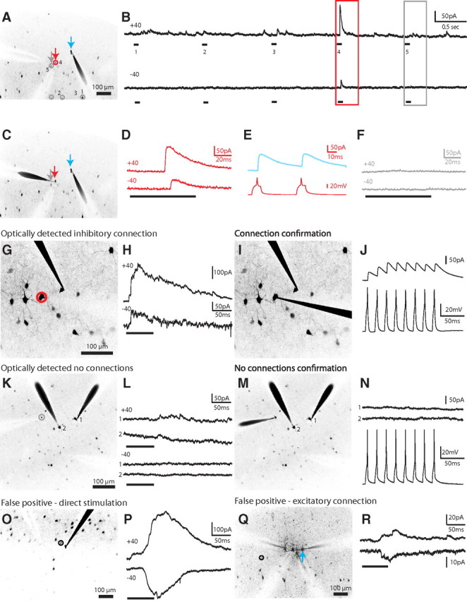
Figure 2.

Mapping inhibitory inputs of pyramidal cells. A, Mapping potential connections from five interneurons (numbered 1 to 5) to one PC (blue arrow) in somatosensory layer 2/3. Gray circles, Unconnected interneurons; red circles, connected. B, Electrophysiological recordings obtained from the PC at holding potentials of +40 mV (top) and −40 mV (bottom) during the photostimulation of the interneurons shown in A. Note the synaptic response from one interneuron (A, red arrow; higher magnification in D) while an interneuron directly nearby (A, gray arrow; higher magnification in F) showed no response. C, E, The red PV+ interneuron neuron was patched and confirmed to be synaptically connected while no response was recorded for the gray neuron (F), which was determined to be unconnected by photostimulation. G, True positive response. A PC patched in a field of view showing many GFP-positive PV+ interneurons nearby, one of which (red circle) was determined to be putatively connected by optical stimulation. H, While holding the PC at +40 mV (top) or −40 mV (bottom), we photostimulated the cell circled in G, eliciting IPSCs. The reversal potential for GABA was set at −80 mV in the internal solution so that EPSCs, but not IPSCs, would change directions between +40 and −40 mV. I, J, The photostimulated cell was patched, confirming electrophysiologically that it was indeed synaptically connected. K, Negative responses. A PV+ interneuron (gray circle) was targeted for photostimulation during whole-cell recording of two nearby PCs. L, The recordings from the PCs in K show no evidence of a synaptic connection during photostimulation of the PV+ interneuron. M, N, The photostimulated cell was patched, confirming electrophysiologically the lack of synaptic connections from the PV+ interneuron onto either PC. O, False positive response. A PV+ interneuron (black circle) directly next to the recorded PC was targeted for photostimulation. P, A slow response, which was outward at +40 mV but flipped at −40 mV, was distinguished from the synaptic event shown in H as a direct stimulation of the patched neuron. Q, A PV+ interneuron (black circle), located distal to the recorded PC (blue arrow), was targeted for photostimulation. R, An excitatory cell connected to the recorded PC was stimulated, resulting in a false positive distinguished by the presence of EPSCs.
