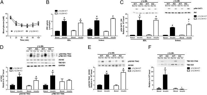
Fig. 3.
AMPK β1β2M-KO mice have normal insulin sensitivity and insulin-stimulated glucose uptake. (A) Whole-body insulin sensitivity in β1β2M-KO mice was assessed by performing an insulin tolerance test (ITT) (0.6 U/kg). (B and C) Insulin-stimulated 2-deoxyglucose (2DG) uptake (B) and Akt S473 phosphorylation (C) in isolated extensor digitorum longus (EDL) and soleus muscles. (D) Insulin-stimulated AS160 T642 and TBC1D1 T596 and phosphorylation in EDL muscle lysates. (E) AS160 T642 phosphorylation in soleus muscle lysates (note TBC1D1 is not detectable in soleus muscle). (F) Insulin-stimulated TBC1D1 PAS phosphorylation in EDL following TBC1D1 immunoprecipitation. Data are means ± SEM, n = 8 ITT and n = 4 ex vivo experiments. *P < 0.05 compared with wild type (WT), same condition. #P < 0.05 compared with basal, same genotype. For protein expression and phosphorylation, values were corrected for equal protein loading using GAPDH or corresponding total antibody.

