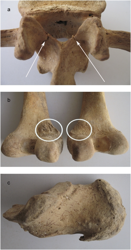
Fig. 4.
Pathological changes in skeletons from Lishui. (A) Dorsal view of the second lumbar vertebra with stress fractures below the articular surfaces on both sides. (B) Dorsal view of both femur condyles. The medial part shows an irregular and porous surface where the attachment of the adductor muscles is located. (C) Lateral view of the right calcaneus. The plantar area is broadened and shows a spur at the attachment of the plantar aponeurosis. Photographs by J.G.

