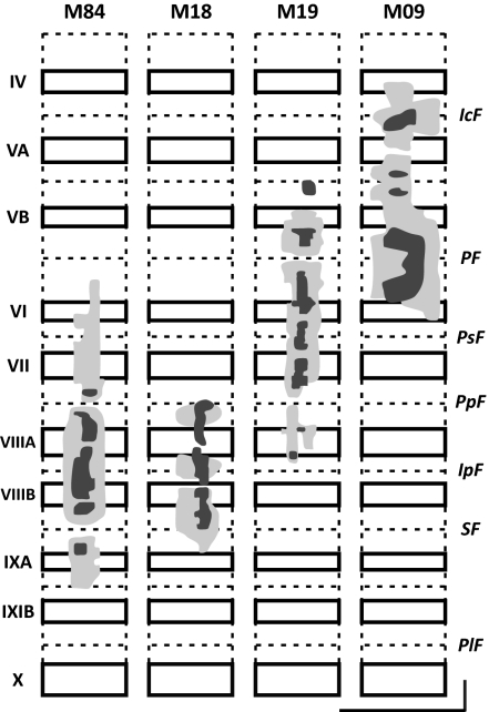
Fig. 2.
Tracer spread in lobules IV–IXA of the vermis. For each lobule, solid rectangles indicate cerebellar cortex on the exposed surface, and dashed lines indicate cerebellar cortex buried in fissures. The injection sites include a dense core (dark shading) and a lighter surround (light shading). All injections were confined to the vermis. (Vertical and horizontal scale bars: 10 mm.) IcF, intraculminate fissure; IpF, intrapyramidal fissure; PF, primary fissure; PlF, posterolateral fissure; PpF, prepyramidal fissure; PSF, posterior superior fissure; SF, secondary fissure.

