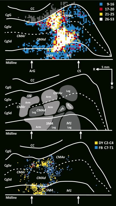
Fig. 7.
Origin of CMAd, CMAv, and SMA projections to the posterior vermis. (Top) Composite map of the labeled neurons in the SMA, CMAd, and CMAv of the seven animals. See Figs. 4 and 6 for conventions. The region of densely labeled neurons (white, yellow, and red bins) overlaps portions of the arm and leg representations within the CMAd, CMAv, and SMA. (Middle) Locations of the arm and leg representations in the motor areas on the medial wall of the hemisphere (adapted from fig. 8 in ref. 12). (Bottom) Location of corticospinal neurons in the motor areas on the medial wall of the hemisphere. Bins indicate the location of neurons labeled by retrograde transport of conventional tracers from upper (yellow) and lower (blue) cervical segments of the spinal cord (adapted from fig. 13 in ref. 12).

