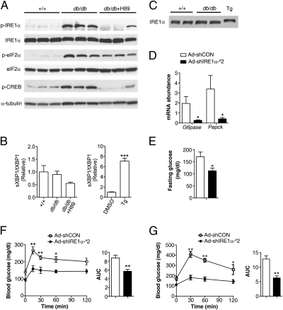
Fig. 5.
PKA-dependent hyperactivation of hepatic IRE1α contributes to obesity-associated disruption of glucose metabolism. (A–C) Highly increased IRE1α phosphorylation in the livers of db/db mice is PKA-dependent and distinct from that induced by ER stress. Male db/db mice were treated for 2 h with PBS or H89 (5 mg/kg body weight) through intraperitoneal injection. For ER stress control, primary hepatocytes were treated with 1 μM Tg or DMSO for 1 h. (A) Liver extracts from db/db mice and their wild-type littermates (+/+) were analyzed by immunoblotting with the indicated antibodies. Shown are representative results for three individual mice from each group (n = 4–5 per group). (B) Abundance of the spliced (s) and total (t) Xbp1 mRNA was determined by real time RT-PCR. (C) Band-shift immunoblot analysis of hepatic IRE1α from wild-type or db/db mice and from Tg-treated hepatocytes. (D–G) Knockdown of hepatic IRE1α improved glucose metabolism in db/db mice. Male db/db mice were infected with Ad-shCON or Ad-shIRE1α-#2 (n = 5/group). (D) At 21 d after infection, the mRNA of liver G6pase and Pepck was analyzed by real-time RT-PCR after a 6-h fast, using actin as an internal control. (E) Glucose was measured after a 6-h fast from mice infected for 18 d. (F and G) Pyruvate tolerance test and glucose tolerance test. Mice infected for 15 or 18 d were fasted for 6 h before intraperitoneal injection with 2 g/kg pyruvate (F) or 1.5 g/kg glucose (G). Blood glucose was measured at the indicated time points, and the AUCs are shown as the mean ± SEM (n = 5/group). *P < 0.05, **P < 0.01 by two-way ANOVA or t test.

