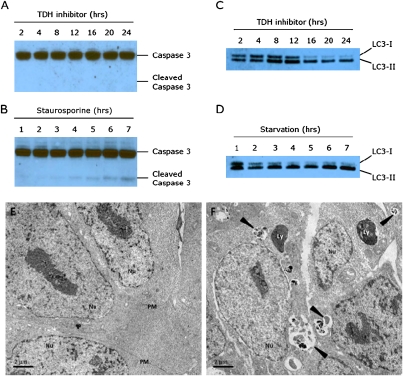
Fig. 5.
TDH inhibition results in elevated autophagy but not apoptosis. (A) Protein lysates from ES cells treated with 10 μM TDH inhibitor were extracted in 0.5% Nonidet P-40, separated using SDS/PAGE, and immuoblotted using an antibody specific for caspase 3. Despite visible cell death, no cleaved caspase 3 could be detected after 24 h of TDH inhibitor treatment. (B) Control Western blot showing cleaved caspase 3 in response to treatment with 1 μM staurosporine. (C) ES cell lysates were immunoblotted with an antibody to LC3. After 16 h of TDH inhibitor treatment, most of the LC3 protein was in the LC3-II (lipidated) form, indicative of increased autophagic activity. (D) Control Western blot showing increased autophagy in response to nutrient starvation. ES cells were cultured in HBSS and harvested at the times indicated. (E and F) Electron microscopy of ES cells treated with TDH inhibitor. Mouse ES cells were cultured on plastic coverslips for 24 h with or without TDH inhibitor. Cells were fixed with 2.5% glutaraldehyde in 0.1 M cacodylate buffer and embedded in Embed-812 Resin. Thin sections (70–90 nm in thickness) were stained with 2% aqueous uranyl acetate and lead citrate and examined by transmission electron microscopy. (E) Control ES cells treated with DMSO only. (F) ES cells cultured in the presence of 10 μM TDH inhibitor. Arrowheads indicate autophagic compartments. Nu, nucleus; PM, plasma membrane; Ly, dense lysosomes.

