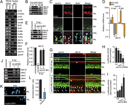
Fig. 1.
Ascl1a inhibits the expression of dkk genes during retina regeneration. (A and B) Injury-dependent regulation of Wnt signaling component mRNAs. (C) Double in situ hybridization shows mutually exclusive ascl1a and dkk1b gene expression. (D) ascl1a and dkk1b expression in FACS-purified MG and non-MG from injured retinas. Values are relative to uninjured retina. *P < 0.009. (E and F) Ascl1a knockdown prevents injury-dependent dkk gene suppression. (F) Quantification of E by qPCR. Values are relative to uninjured retina. *P < 0.0001. (G) In situ hybridization showing Ascl1a knockdown relieves injury-dependent dkk suppression. Boxed region in low-magnification image is shown in higher magnification in the row below. Arrows point to ascl1a+/dkk1b+ cells. White dots identify autofluorescence in ONL. (H and I) Injection of zebrafish embryos with dkk1b:gfp-luciferase reporter and increasing amounts of ascl1a mRNA (H) or ascl1a-targeting MO (I). *P < 0.005. (J) Lin-28 knockdown differentially affects injury-dependent dkk gene suppression. (K and L) Dkk1b overexpression inhibits cell proliferation at 4 dpi. *P < 0.003. (Scale bars, 10 μm.) ONL, outer nuclear layer; INL, inner nuclear layer; GCL, ganglion cell layer.

