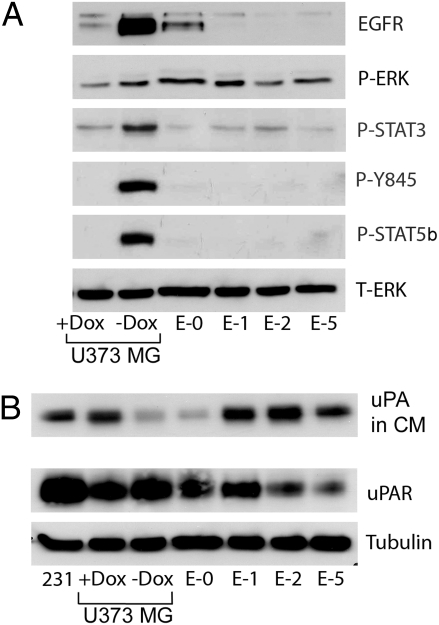
Fig. 1.
Tyr-845 and STAT5b are phosphorylated in EGFRvIII-expressing U373MG cells. (A) U373MG cells were treated with Dox (1 μg/mL) or vehicle for 5 d and maintained in SFM for the last 2 d. Escaper tumor cells (E-0, E-1, E-2, and E-5) were maintained in Dox and treated similarly. Cell extracts were subjected to immunoblot analysis to detect P-ERK, P-STAT3, P–Tyr-845 in the EGFR, P-STAT5b and total ERK (T-ERK). (B) Conditioned SFM (CM) was collected from cultures at equivalent confluency and concentrated 10 times. Immunoblot analysis was performed to detect uPA. The control lane labeled “231” shows CM from MDA-MB-231 cells, which express high levels of uPA (29). Cell extracts were subjected to immunoblot analysis to detect uPAR and tubulin as a loading control.

