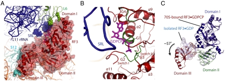
Fig. 4.
Conformation of the ribosome-bound GTPase RF3 in translation. (A) Conformation of RF3•GDPCP in the ribosome, with the interacting residues shown as spheres. (B). The sarcin-ricin loop in the 23S rRNA is in the proximity to the nucleotide binding pocket of RF3. GTP substrate analogue GDPCP is colored in magenta, and the four G motifs in the RF3 are highlighted in forest. (C). Conformational changes of RF3 upon ribosome binding. The ribosome-bound RF3•GDPCP (colored by individual domains as domain I in blue, domain II in green, and domain III in firebrick) adopts a different conformation compared to the isolated RF3•GDP structure (light blue) (8).

