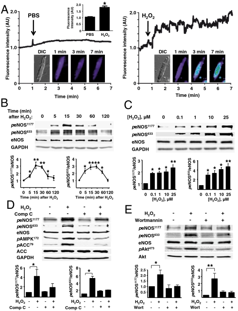
Fig. 1.
Effects of H2O2 on cardiac myocyte NO synthesis and eNOS phosphorylation. (A) Adult mouse cardiac myocytes were loaded with the NO dye Cu2(FL2E), and then treated either with PBS or H2O2 (10 μM). Fluorescence tracings are shown from a typical experiment, as well as representative differential interference contrast (DIC) and fluorescence images (1, 3, and 7 min). AU, arbitrary units. (B and C) Representative immunoblots from time course (B) or dose-response (C) experiments documenting the effects of H2O2 on eNOS phosphorylation at Ser1177 (peNOS1177) or Ser633 (peNOS633). (D) Cardiac myocytes were incubated with compound C (Comp C, 20 μM, 30 min) or vehicle, then treated with H2O2 and analyzed in immunoblots probed with antibodies as shown. (E) Immunoblot analyses from cardiac myocytes incubated with the PI3-kinase inhibitor wortmannin (1 μM, 30 min) or vehicle, then treated with H2O2. Below each representative immunoblot are shown the results of densitometric analyses from pooled data, documenting the changes in peNOS1177 and peNOS633 plotted relative to the signals present in unstimulated cells. Each data point represents the mean ± SE derived from at least three independent experiments; * indicates p < 0.05 and ** indicates p < 0.01.

