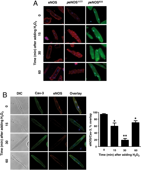
Fig. 2.
H2O2-promoted eNOS phosphorylation and translocation in cardiac myocytes. (A) Results of immunohistochemical analyses of cardiac myocytes that were treated with H2O2 (10 μM) for the indicated times, and then fixed, permeabilized, probed with antibodies against total eNOS, peNOS Ser1177, or peNOS Ser633, and imaged using confocal microscopy. (B) Images of cardiac myocytes treated with H2O2 (10 μM) for the indicated times and then fixed, permeabilized, and probed with antibodies against total eNOS (Alexa Fluor red 568) or Cav-3 (Alexa Fluor green 488); overlapping signals are shown in yellow. The images shown on the left are representative of three independent experiments that yielded similar results; the bar graph on the right shows pooled data from three experiments, quantitating the percent overlap between eNOS and Cav-3 at different times after adding H2O2; * indicates p < 0.05; ** indicates p < 0.01 compared to t = 0.

