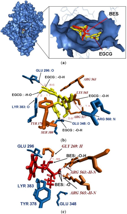
Figure 3.
Micro environment of the bind site of EGCG/BES and Leukotriene A4 hydrolase. (a) EGCG (yellow) binds to Leukotriene A4 hydrolase in the active cavity (catalytic center and binding domain) where the original ligand BES (red) located; (b) Residues GLU348, SER380, TYR383, TYR378, ARG563, ARG565, ARG568, GLU296 and ARG537 make up a electrostatic cavity; hydroxyl groups of EGCG form hydrogen bonds with the surrounding amino acids GLU348, TYR383, GLU296 and ARG568 (blue); (c) The original ligand formed 3 strong hydrogen bonds with residues GLY269, ARG563, and ARG565 (orange).

