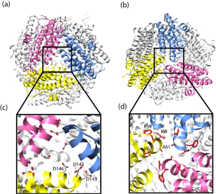
Figure 5.
DPS mini-ferritin protein cages viewed along the (a) ferritin-like three-fold axis; and (b) Dps-like three-fold axis; (c) The expansion shows the aspartic acid residues lining the ferritin-like pore; (d) The expansion shows the hydrophobic amino acids lining the DPS-like pore. Note: Due to symmetry, the residues in (c) and (d) are labeled on only one monomer. The figure is generated using UCSF Chimera [44] (PDB ID: 1 dps).

