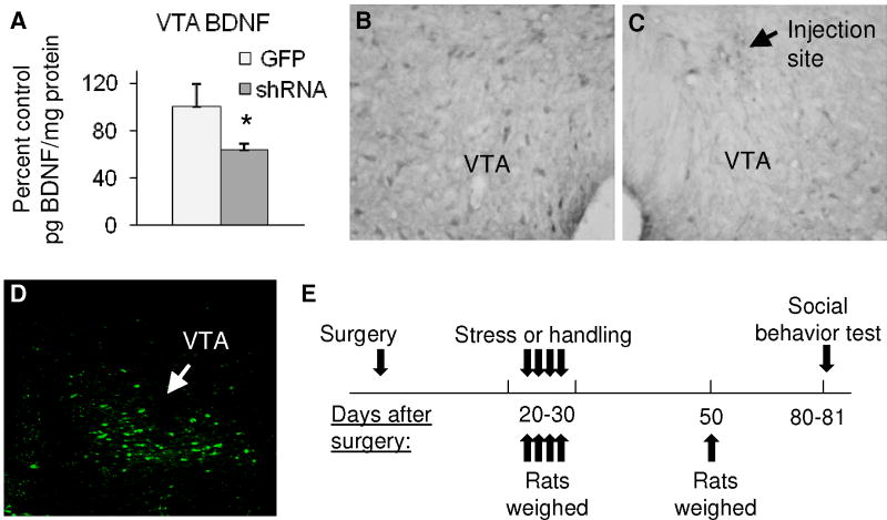
Figure 1. AAV-mediated depletion of BDNF in the VTA and timeline of experiments.
A - Percent control BDNF expression (pg BDNF/mg protein). Infusion of AAV-GFP-shRNA into the VTA reduced BDNF by approximately 36% (*p < 0.05; n = 6–10) as measured by BDNF ELISA in animals euthanized 3 weeks after viral infusion. B - BDNF immunolabeling in the intact VTA and C - near the injection site of AAV-GFP-shRNA 3 weeks earlier (contralateral side of the same section as shown in B); magnification 200X. D - Fluorescence image of GFP expression in rat VTA 3 weeks after viral infusion; magnification 100X. E -Timeline of behavioral experiments.

