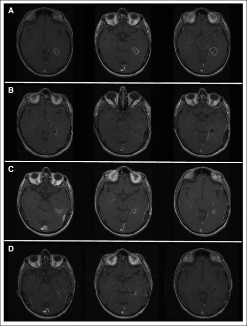
Fig 3.
Slow resolution of magnetic resonance imaging enhancement; magnetic resonance imaging of patient 1B3 at dose level 1. Before surgery and injection (panel A) imaging shows a glioblastoma multiforme in the left temporal lobe. Postoperative images (panel B) reveal a total resection. Residual ring enhancement is apparent 4 weeks after completion of radiation (panel C), which gradually resolved over the next 6 months. Decreased enhancement 4 months after radiation completion is shown in panel D. Adjuvant temozolomide was stopped after four cycles because of noncompliance. However, the patient did not develop progression until 27 months and survived for 46 months.

