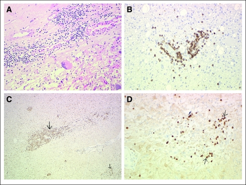
Fig 4.
Histologic analysis of re-resected tumors. Re-resection of glioblastoma multiforme at 30 weeks in patient 1B4 with demonstration of intratumoral lymphocytic infiltrate (A) via staining with hematoxylin and eosin, which was found to be CD3+ T cells by immunohistochemistry. (B) Anti-CD3 antibody. Re-resection of glioblastoma multiforme at 36 weeks in patient 2B2 with demonstration of macrophage (C) and cell (D) infiltration. (C) Anti-CD68 antibody. (D) Anti-CD3 antibody. Anti-CD20 staining for B cells was negative (not shown). Arrows point to positive cells.

