
Fig. 1.
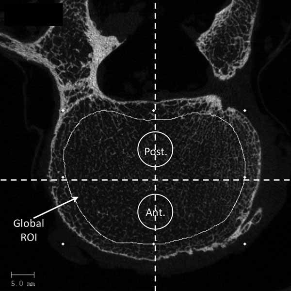
HR-pQCT slice of L3 vertebra. Trabecular region of interest (ROI) was defined manually in order to exclude cortical component of the vertebral body. Virtual biopsies were positioned using two lines drawn on the vertebral body, one line for the middle anteroposterior axis and one line for the middle mediolateral axis. Each line divided the vertebral body into four quadrants. Biopsies were strictly centered on the middle anteroposterior axis and on both sides of the mediolateral axis to avoid the cortical shell anteriorly and the venus plexus posteriorly by projection in the vertical direction in the HR-pQCT slice stack.