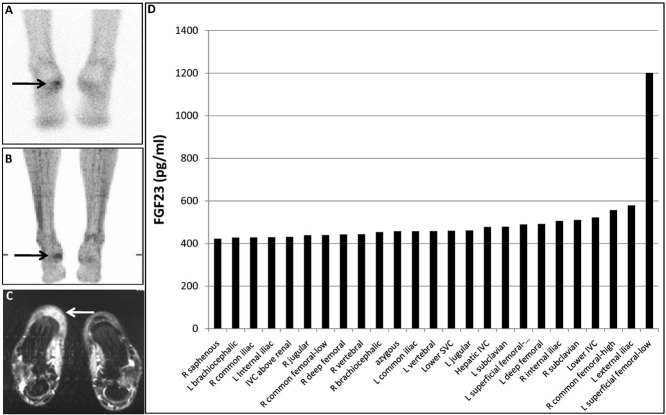
Fig. 1.
Example of a positive result on selective venous sampling. A suspicious lesion was identified by indium-111 pentetreotide scintigraphy (A) and FDG-PET scan (B) that correlated with an ill-defined lesion in the fat pad of the heel on MRI (C). Surgical removal of the fat pad would entail an extensive procedure that involved the translocation of a vascularized muscle flap from the arm. The results of the selective venous sampling measurements of FGF-23, which confirmed that the lesion was the FGF-23-secreting lesion, are shown (D) The tumor is indicated by the arrows.

