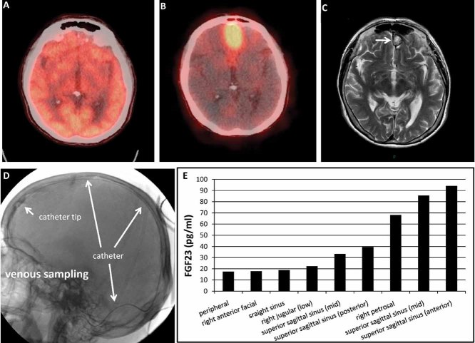
Fig. 4.
Example of a selective venous sampling that distinguished between conflicting imaging results. For a lesion that ultimately was found adjacent to the brain, FDG-PET/CT scan (A) was negative owing to the intense physiologic uptake of FDG by the brain. The pentetreotide/CT scan was positive (B), but the MRI findings (C) were felt to be most consistent with a meningioma, which also takes up pentetreotide. To resolve the matter, the superior sagittal sinus was catheterized from a transjugular approach (D), and FGF-23 concentrations were determined (E). The catheter and catheter tip are identified as indicated by white arrows. The results of the venous sampling were consistent with the fact that the lesion identified on pentetreotide scan and MRI was the FGF-23-secreting tumor.

