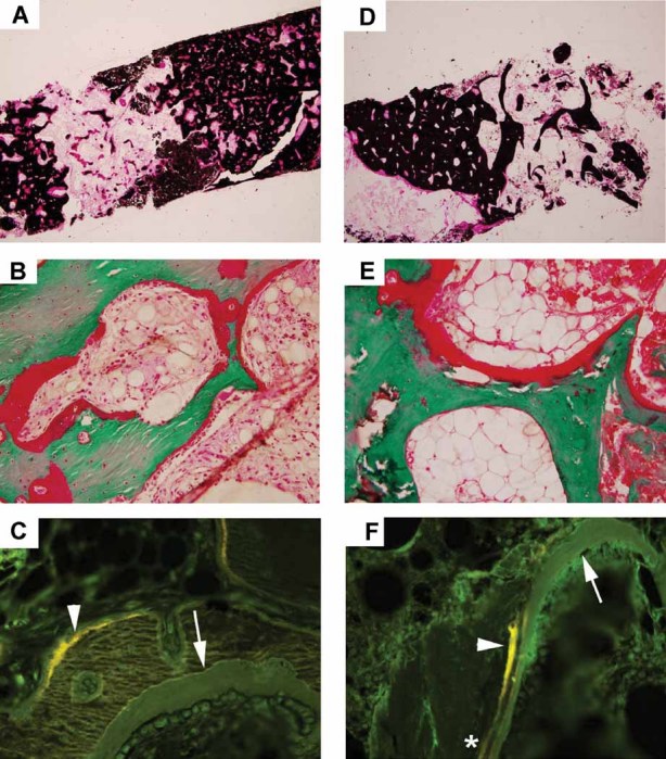
Fig. 5.

Histological findings: (A-C) The iliac crest specimens from patient 1 (son) and (D–F) patient 2 (mother). Overall, the findings were consistent with osteomalacia that was more striking in the son. (A,D) Von Kossa–stained sections show thickened porous cortices and decreased trabecular connectivity. (B,E) Goldner trichrome–stained sections show thick osteoid seams covering most trabecular and cortical surfaces. (C,F) Fluorescence microscopy shows broad single tetracycline labels (arrowheads) as well as unlabeled osteoid (arrows). Although double tetracycline labels could be found in both patients, they were more numerous in the mother (asterisk).
