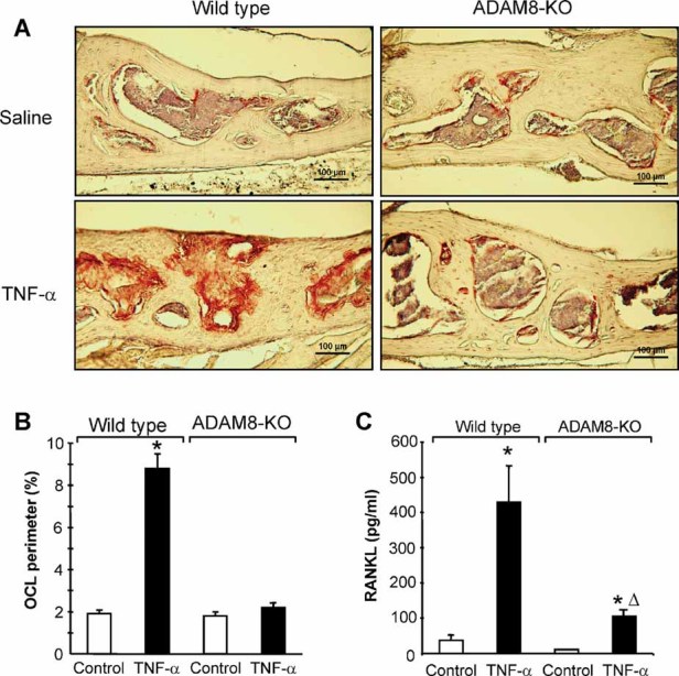
Fig. 7.

Responses to TNF-α administration in ADAM8 KO and WT mice. Photomicrographs taken from sections of calveria from WT and ADAM8 KO mice treated with saline or TNF-α and stained with TRAP and hematoxylin. WT mice demonstrated increased osteclastogenesis, as demonstrated by the increased numbers of TRAP+ (red) osteoclasts present in deep resorption cavities and inflammation. These responses were blunted in ADAM8 KO mice treated with TNF-α (p < .01). (B) OCL perimeter measured in WT and ADAM8 KO mice treated with TNF-α or saline. TNF-α significantly increased OCL perimeter in WT but not ADAM8 KO mice. *Significantly different from saline treatment (p < .01). (C) RANKL production by marrow stromal cells from ADAM8 KO and WT mice. Marrow-adherent cells were isolated and cultured for 2 days with vehicle or TNF-α (10 ng/mL). The conditioned media were harvested and RANKL levels measured with an ELISA assay from R&D Systems (Minneapolis, MN, USA). Results expressed the mean ± SD for four determinations. *Significantly different from saline-treated cultures (p < .01). ΔSignificantly different from WT cultures treated with TNF-α.
