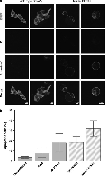
Figure 3.
(a) Confocal images of annexin V-Cy5-stained cell transfections. Cells transfected with WT DFNA5 are negative for PI and annexin V-Cy5. Cells transfected with mutant DFNA5 are negative for PI, but show clear annexin V at the plasma membrane, which is suggestive for apoptotic cell death. All cells were harvested 16 h post-transfection. A full color version of this figure can be found in the html version of this paper. (b) Flow cytometric quantification of annexin V-Cy5-stained transfected cells. Cells were harvested 16 h post-transfection. Percentages of apoptotic cells within the total cell population are shown. The total apoptotic fraction was calculated as the sum of the early (annexin V+ PI−) and late (annexin V+ PI+) apoptotic fractions.

